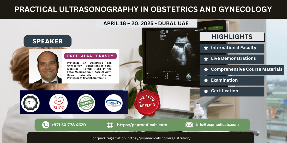

Practical Ultrasonography In Obstetrics And Gynecology
OVERVIEW
Practical Ultrasonography in Obstetrics and Gynecology, organized by PSP Medicals, will take place from April 18-20, 2025, at the Grand Hyatt Dubai, United Arab Emirates.
Description:
Enhance your diagnostic skills in obstetric and gynecological ultrasonography through this hands-on workshop. Learn from expert instructors and gain practical experience using state-of-the-art ultrasound equipment during live demonstrations and practice sessions.
What’s Included:
- Live Demonstrations & Hands-On Training:
Each session will begin with a live demonstration followed by hands-on practice to help you develop essential skills in performing ultrasound examinations in obstetrics and gynecology. - DHA CME Credits:
The program is accredited for Continuing Medical Education (CME) by the Dubai Health Authority (DHA), contributing to your professional development. - Diploma Certificate:
Upon successful completion, participants will receive the prestigious Diploma in Practical Ultrasonography in Obstetrics and Gynecology, accredited by the International Society of Ultrasound in Obstetrics and Gynecology (ISUOG). - Comprehensive Course Materials:
Receive detailed materials, protocols, and notes to support your learning and practical application of ultrasonography techniques. - Accommodation:
Enjoy 3 Nights of Free Accommodation at a luxurious 5-star hotel during the event. - Visa Assistance:
Visa assistance will be provided upon request for international participants.
Why Attend?
- Hands-On Learning: Gain real-world experience with live cases and hands-on practice using ultrasound technology in obstetrics and gynecology.
- International Accreditation: Earn a diploma accredited by ISUOG, a globally recognized institution in ultrasound education.
- Enhance Your Skills: Learn to perform essential ultrasound procedures for obstetric and gynecological diagnoses with confidence.
- CME Credit: Enhance your professional qualifications with DHA CME credits for this valuable training.
COURSE CONTENTS
- Fetal Anatomy Scan from A to Z
A systematic approach to the detailed anomaly scan (18–22 weeks), covering:- Comprehensive evaluation of fetal organ systems:
- Central nervous system
- Cardiovascular system
- Abdominal organs and skeletal structures
- Placenta, amniotic fluid, and umbilical cord assessment
- Common anomalies and diagnostic protocols
- Practical tips for accurate image acquisition
- Comprehensive evaluation of fetal organ systems:
- Color Doppler in Obstetrics: How and When
Understanding Doppler ultrasound principles and applications:- Key indications:
- Trophoblastic flow in the first trimester
- Umbilical artery, middle cerebral artery, and ductus venosus evaluation
- Doppler use in high-risk pregnancies:
- Fetal growth restriction
- Preeclampsia monitoring
- Optimizing machine settings for accurate measurements
- Troubleshooting common Doppler artifacts
- Key indications:
- The 13-Week Scan: A Paradigm Shift in Fetal Medicine
Early pregnancy screening and diagnostic approaches at 11–13+6 weeks, including:- Key markers:
- Nuchal translucency, nasal bone, and ductus venosus flow
- Integration with biochemical markers (PAPP-A, free beta-hCG)
- Early detection of structural anomalies (CNS, abdominal wall defects)
- Placental and uterine artery Doppler for preeclampsia risk assessment
- Implications for personalized pregnancy management
- Key markers:
- How to Write a Gynecological Ultrasound Report
Key components of a structured and comprehensive report:- Patient history and clinical indications
- Detailed ultrasound findings: uterus, ovaries, adnexa, and endometrium
- Standardized terminology and classifications (e.g., IOTA)
- Common reporting errors and how to avoid them
- Legal and ethical aspects of report documentation
- Twins by Ultrasound
Determining chorionicity and amnionicity in early pregnancy:- Monitoring growth and complications in twin pregnancies:
- TTTS, TAPS, sIUGR, and TRAP sequence
- Biometric discordance and implications for management
- Anomaly detection in multiple pregnancies
- Delivery planning based on chorionicity and other factors
- Monitoring growth and complications in twin pregnancies:
- 3D/4D Ultrasound in Obstetrics and Gynecology
Principles of volume acquisition and rendering techniques:- Applications in obstetrics:
- Fetal face and spine assessment
- Congenital anomaly visualization
- Applications in gynecology:
- Uterine anomalies and infertility evaluation
- Endometrial assessment and adnexal pathology
- Integration of advanced imaging into clinical practice
- Future trends and emerging technologies
- Applications in obstetrics:
KEY DATES
- Registrations Open: December 28, 2024
- Registration Ends: March 31, 2025
- Event Start Date: April 18, 2025
- Event End Date: April 20, 2025
INTERESTED PARTICIPANTS
- Course – Early Fee: US$2,000.00
- Registration Closes: March 31, 2025
SPEAKERS
- Alaa El Din Nagiub El Ebrashy, MD, Obstetrics and Gynecology, Maternal Fetal Medicine
TARGET AUDIENCE
- Physicians, Nursing Professionals, Physician Assistants, Sonographers, Obstetrics and Gynecology Physicians, Healthcare Professionals, Nurses, Paediatric Urology, Obstetricians, Ultrasound Specialists, Gynecologists, Ultrasonographers, Gynecological Oncologists
SPECIALITIES
- Obstetrics and Gynecology
- Sonography
- Female Pelvic Medicine and Reconstructive Surgery
Entry Requirements
- Medical Doctors (MD, MBBS, DO)
- Nurses and Nurse Practitioners
- Physician Assistants
- Healthcare Professionals
Fees
- Diploma Certificate: USD 2,500
- Early Bird Discount: USD 500 (valid until March 31, 2025)
- 3 Nights Free Accommodation: Included in a 5-star hotel
- Visa Assistance: Provided upon request
Please proceed with the partial payment of USD 1,000 (AED 3,600) via the provided link. The remaining balance can be settled on-site.
Course Materials
- All course materials will be provided.
- Each session will begin with a live demonstration, followed by hands-on practice to ensure practical skill development in Ultrasonography.
Certificate
Upon successful completion of the program, you will receive the prestigious Diploma in Practical Ultrasonography in Obstetrics and Gynecology, accredited by the International Society of Ultrasound in Obstetrics and Gynecology (ISUOG). Participants will also receive comprehensive course materials, protocols, and detailed notes to support their learning and application of ultrasonography techniques.


